There is a third difference between cameras and the human visual system (HVS). While some camera lenses may share a similar perspective of the world with the HVS with respect to the angle-of view, where they differ is what is actually in the area of focus. Using any lens on a camera means that a picture will have an area where the scene is in-focus, with the remainder being out-of-focus. This in-focus region generally occurs in a plane, and is associated with the depth-of-field. On the other hand, the in-focus region of the picture our mind presents us does not have a plane of focus.
While binocular vision allows approximately 120° of (horizontal) vision, it is only highly focused in the very centre, with the remaining picture being increasingly out-of-focus depending on how far a point is away from the central focused region. This may be challenging to visualize, but if you look at an object, only the central point is in focus, the remainder of the picture is out-of-focus. That does not mean it is necessarily blurred, because the brain is still able to discern shape and colour, just not fine details. Blurring it usually a function of distance from the object being focused on, i.e. the point-of-focus. If you look at a close object, distant objects will be out-of-focus, and vice versa.
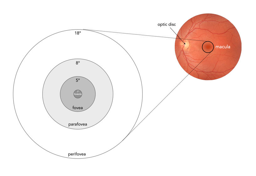
Focused vision is related to the different parts of the macula, an oval-shaped pigmented area in the centre of the retina which is responsible for interpreting vision, colour, fine details, and symbols (see Figure 1). It is composed almost entirely of cones, into a series of zones:
- perifovea (5.5mm∅, 18°) : Details that appear in up to 9-10° of visual angle.
- parafovea (3mm∅, 8°) : Details that appear in peripheral vision, not as sharp as the fovea.
- fovea (1.5mm∅, 5°) : Or Fovea centralis, comprised entirely of cones, and responsible for high-acuity, and colour vision.
- foveola (0.35mm∅, 1°) : A central pit within the fovea, which contains densely packed cones. Within the foveola is a small depression known as the umbo (0.15mm∅), which is the microscopic centre of the foveola.

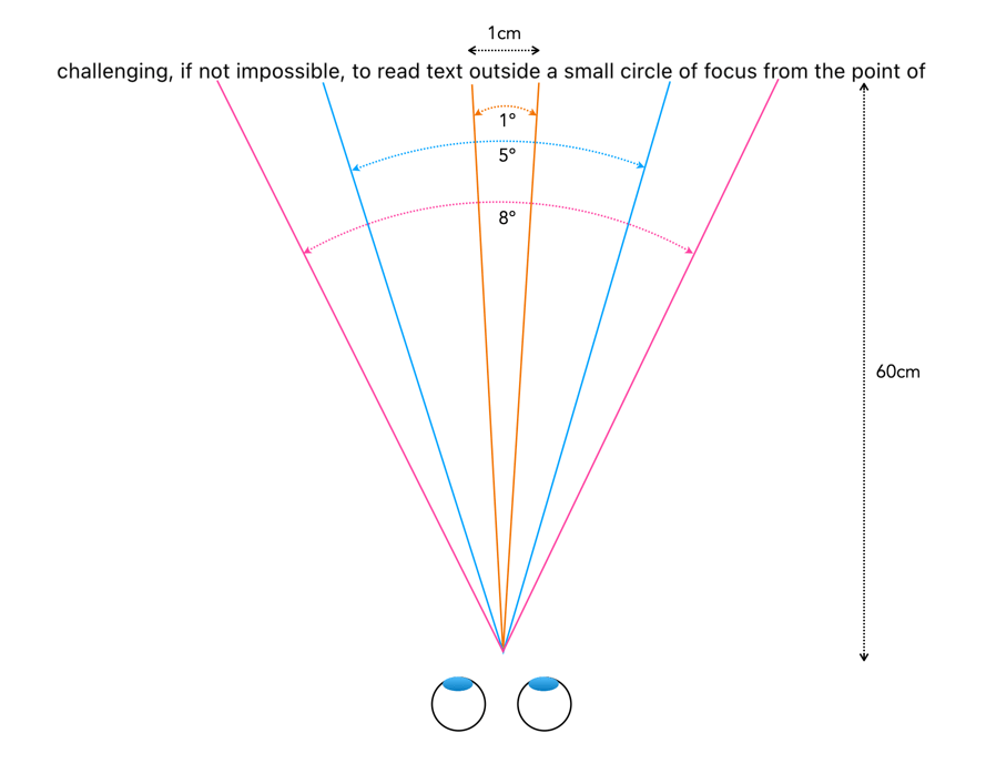
When we fixate on an object, we bring an image of that object onto the fovea. The foveola provides the greatest amount of visual acuity, in the area 1-2° outwards from the point of fixation. As the distance from fixation increases, visual acuity decreases quite rapidly. To illustrate this effect, try reading the preceding text in this paragraph while fixating on the period at the end of the sentence. It is likely challenging, if not impossible, to read text outside a small circle of focus from the point of fixation. A seven letter word, like “outside”, is about 1cm wide, which when read on a screen 60cm from your eye represents about an angle of 1°. The 5° of the fovea region allows for a “preview” of the words either side, and parafovea region, 8° of peripheral words (i.e. their shape). This is illustrated in Figure 3.
To illustrate how this differential focus affects how humans view a scene, consider the image shown in Figure 4. The point of focus is a building in the background roughly 85m from where the person is standing. This image has been modified by adding radial blur from a central point-of-focus to simulate in-focus versus out-of-focus regions as seen by the eye (the blur has been exaggerated). The sharpest region is the point of fixation in the centre – from this focus on a particular object, anything either side of that object will be unsharp, and the further away from that point, the more unsharp is becomes. The
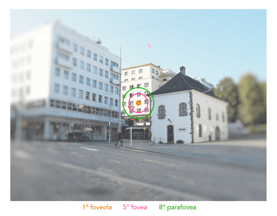
It is hard to effectively illustrate exactly how the HVS perceives a scene as there is no way of taking a snapshot and analyzing it. However we do know that focus is a function of distance from the point-of-focus. Other parts of an image as essentially de-emphasized, there is still information there, and the way our minds process it, it provides a complete vision, but there is a central point of focus.
Further reading:
- Ruch, T.C., “Chapter 21: Binocular Vision, and Central Visual Pathways”, in Neurophysiology (Ruch, T.C. et al. (eds)) p.441-464 (1965)


