In the previous post, from a pure pixel viewpoint, we got a mixed bag of numbers to represent the human eye in terms of megapixels. One of the caveats was that not all pixels are created equal. A sensor in a camera has a certain resolution, and each pixel has the same visual acuity – whether a pixel becomes sharp or blurry is dependent on characteristics such as the lens, and depth-of-field. The human eye does not have a uniform acuity.
But resolution is about more than just how many pixels – it is about determining fine details. As noted in the last post, the information from the rods in coupled together, whereas the information from each cone has a direct link to the ganglion cells. Cones are therefore extremely important in vision, because without them we would view everything as we do in our peripheral vision, oh and without colour (people who can’t see colour have a condition called achromatopsia).
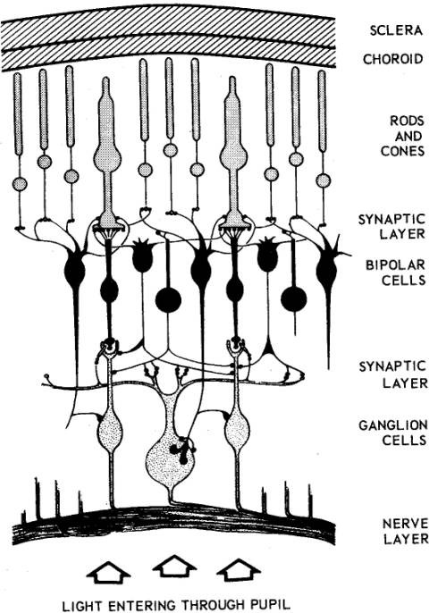
Cones are however, not uniformly distributed throughout the retina – they are packed more tightly in the centre of the eyes visual field, in a place known as the fovea. So how does this effect the resolution of the eye? The fovea (which means pit), or fovea centralis, is located in the centre of a region known as the macula lutea, a small oval region located exactly in the centre of the posterior portion of the retina. The macula lutea is 4.5-5.5mm in diameter, and the fovea lies directly in the centre. The arrangement of these components of the retina is shown below.

The fovea has a diameter of 1.5mm (although it varies slightly based on the study), and a field of view of approximately 5°. Therefore the fovea has an area of approximately 1.77mm². The fovea has roughly 158,000 cones per mm ² (see note). The density in the remainder of the retina is 9,000 cones per mm². So, the resolution of the human eye is much greater in the centre, than on the periphery. This high density of cones is achieved by decreasing the diameter of the cone outer segments such that foveal cones resemble rods in their appearance. The increased density of cones in the fovea is accompanied by a decrease in the density of rods. Within the fovea is a region called the foveola which has a diameter of about 0.3mm, and a field of view of 1° – this region contains only cones. The figure below (from 1935) shows the density of rods and cones in the retina.

The fovea has the highest visual acuity in the eye. Why? One reason may be the concentration of colour-sensitive cones. Most photoreceptors in the retina are located behind retinal blood vessels and cells which absorb light before it reaches the photoreceptor cells. The fovea lacks the supporting cells and blood vessels, and only contains photoreceptors. This means that visual acuity is sharpest there, and drops significantly moving away from this central region.
For example pick a paragraph of text, and stare at a word in the middle of it. The visual stimulus in the middle of the field of view falls in the fovea and is in the sharpest focus. Without moving your eye, notice that the words on the periphery of the paragraph are not in complete focus. The images in the peripheral vision have a “blurred” appearance, and the words cannot be clearly identified (although we can’t see this properly, obviously). The eyes receive data from a field of view of 190-200°, but acuity of most of that range is quite poor. If you view the word from approximately 50cm away, then the field of view is about ±2.2cm from the word – beyond that things get fuzzier. Note that each eye obviously has its own fovea, but when you focus on a point both fovea overlap, but the resolution doesn’t increase.
The restriction of highest acuity vision to the fovea is the main reason we spend so much time moving our eyes (and heads) around. From a processing perspective, the fovea represents 1% of the retina, but the brains visual cortex devotes 50% of its computation to input from the fovea. So in the fovea, resolution is equivalent of a TIFF, whereas elsewhere it’s a JPEG. So, if the sharpest and most brilliantly coloured human vision comes from the fovea, what is its resolution?
Again this is a somewhat loaded question, but let’s attempt it anyway. If the fovea has a field of view of 5°, and assuming a circular region, we can create a circular region with a radius 2.5 degrees = 19.635 degrees2, and 60×60 = 3600 arcmin2/degree2. Assume a “pixel” acuity of 0.3×0.3=0.09 arcmin2. This gives us 19.635*3600 / 0.09 = 785,400 pixels. Even if we round up we get a resolution of about 1MP for the fovea. And honestly, the actual point of highest acuity may be even smaller than that – if we considered the foveola, we’re looking at a mere 125,000 pixels.
NOTE
Note: There are many studies relating to the size of the fovea, and the density of photoreceptors, given that each human is a distinct being, there is no one exact number.
Jonas, J.B., Schneider, U., Naumann, G.O.H., “Count and density of human retinal photoreceptors”, Graefe’s Archive for Clinical and Experimental Ophthalmology, 230(6), pp.505-510 (1992).
For more information on the anatomy of the retina.
Everything you wanted to know about visual acuity.
